Analytical investigation of metal distribution from e-cigarette aerosols to lung deposition using multi-platform mass spectrometry
Anal Bioanal Chem 418, 3821–3835 (2026): Graphical abstract
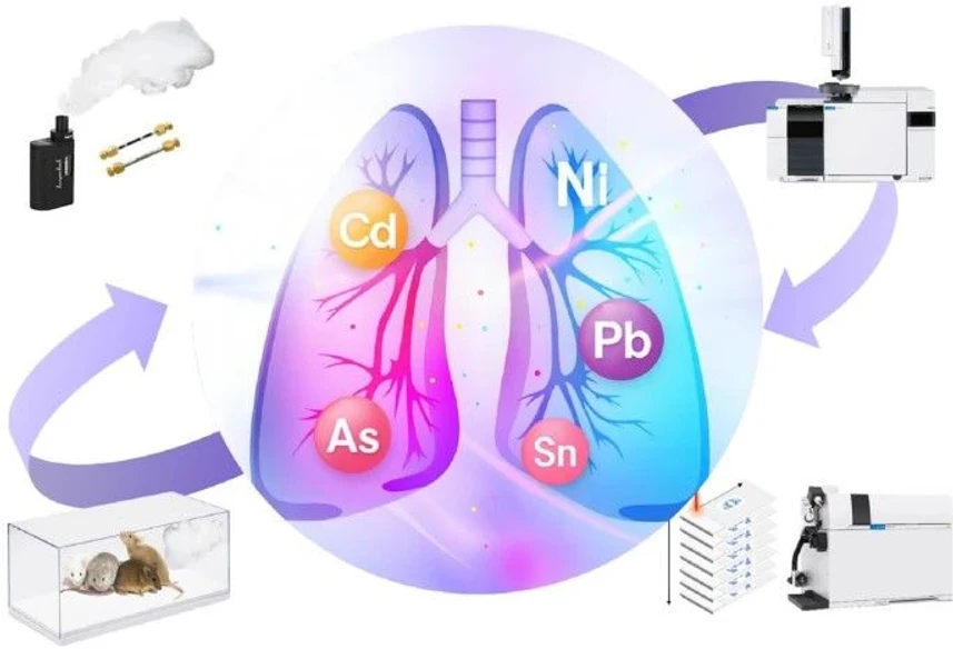
This study investigates the transfer of metals from e-cigarette liquids and aerosols into lung tissue using a combination of ICP-MS, GC-ICP-MS, GC-MS, and LA-ICP-MS/MS. Multiple toxic metals and organometallic species, including aluminum, nickel, copper, arsenic, tin, and mercury, were detected in both e-liquids and generated aerosols.
Exposure experiments in mice revealed heterogeneous accumulation of metals within lung tissue, with significant increases in nickel and lead and decreased iron levels. The findings provide the first evidence of organometallic species in e-cigarette aerosols and demonstrate exposure-related metal deposition in the lungs, highlighting the need for further research into the long-term health effects of vaping.
The original article
Analytical investigation of metal distribution from e-cigarette aerosols to lung deposition using multi-platform mass spectrometry
Jack McGrath, Oliver Royle, Andrew Thorpe, Janice Irene McCauley, Maiken Ueland, Irina Kabakova, Hui Chen, David Clases, Brian G. Oliver & Dayanne Mozaner Bordin
Anal Bioanal Chem 418, 3821–3835 (2026)
https://doi.org/10.1007/s00216-026-06487-1
licensed under CC-BY 4.0
Selected sections from the article follow. Formats and hyperlinks were adapted from the original.
Since their introduction in 2004, electronic cigarettes (e-cigarettes, vapes) have been promoted as a safer alternative to traditional tobacco and as an effective tool for smoking cessation aid [1, 2]. These devices deliver inhalable nicotine generated by heating a liquid formulation, a process claimed to avoid the combustion-related by-products such as tar and many carcinogens associated with conventional cigarettes [1, 2]. This perceived reduction in harm, along with misleading marketing campaigns [3, 4], has contributed to the rapid uptake of e-cigarettes globally, particularly among younger demographics. In Australia, for example, e-cigarette use among young adults increased from 5.3% in 2019 to over 21% in 2023, with a similar rise in adolescents [5]. Comparable trends are seen in the USA and the UK, where e-cigarette use among young people has become a significant public health concern [6, 7]. This uptake was partially driven by the availability of unregulated and often illegally imported and low-cost devices, which are sold to children.
The long-term health impacts of e-cigarettes, however, are unclear due to their recent introduction to the market. Nonetheless, inhalation of e-cigarette aerosols has been associated with adverse respiratory outcomes, including chronic obstructive pulmonary disease (COPD), airway inflammation, impaired lung function, and increased oxidative stress, much the same as tobacco [6,7,8]. Unlike cigarettes, which are a relatively consistent global product, e-cigarette formulations and devices are often manufactured with poor quality control, often involving materials and components with unknown toxicological relevance. Limited regulatory oversight further necessitates systematic investigation of the chemical composition of these products and their potential respiratory impacts [7, 8].
E-cigarette aerosols are complex mixtures containing volatile organic compounds (VOCs), aldehydes, tobacco-specific nitrosamines (TSNAs), and metals such as chromium (Cr), nickel (Ni), copper (Cu), and lead (Pb) [9,10,11]. These toxicants may originate from device components, including heating elements, or the e-liquids themselves [9]. Previous studies have reported substantial variability in metal and other chemical concentrations across products, reflecting differences in device design, materials, manufacturing quality, and user behaviour [9, 12, 13]. Elevated levels of metals such as Cr, Cu, and Pb have been detected in biological samples (urine, saliva, serum, and blood) of e-cigarette users compared to non-users [14], while prolonged inhalation exposure to Ni and Pb has been associated with pulmonary inflammation, oxidative stress, and structural alterations in lung tissue in experimental and epidemiological studies [15, 16].
Despite this growing body of evidence, the extent to which metals derived from e-cigarette aerosols accumulate within lung tissue, and their spatial distribution following inhalation, remains poorly characterised. Recent investigations indicate that device components composed of Ni-Cr alloy, stainless-steel, and other internal materials can contribute to metal transfer into e-liquids and aerosols during use, with release influenced by device ageing and coil degradation. Metals such as Cr and Ni are primarily associated with heating elements, whereas Pb, Cu, Zn, and antimony (Sb) may originate from non-heating components. The speciation studies demonstrate that metals are present in defined chemical forms, such as Cr predominantly as Cr(III) and Sb as mixed Sb(III)/Sb(V) species, emphasising the importance of integrating speciation with total elemental analysis when evaluating exposure and potential health implications [17]. This information is essential for assessing the long-term health risks, particularly in vulnerable populations, and for informing regulatory policies.
Toxic metal exposure is a recognised global health concern, with some acting as systemic toxicants even at low levels [18, 19]. Accordingly, there is a need for robust analytical characterisation of metals and metal species associated with e-cigarette use. Therefore, this study investigated whether short-term exposure to e-cigarette aerosols leads to metal accumulation in lung tissue, alongside a systematic characterisation of elemental and metal-containing species in the aerosol. It used a murine inhalation model with complementary mass spectrometric techniques, including high-resolution elemental bioimaging, to quantify tissue-associated metals and determine their spatial distribution within the lung.
Materials and methods
Instrumentation, experimental parameters, and sample preparation
A multi-platform analytical approach was employed to characterise the elemental and organic composition of e-cigarette liquid and to assess the presence of metal-containing species in the corresponding aerosol, as well as to evaluate metal accumulation in lung tissue following the exposure protocol. Instrumental parameters were systematically optimised prior to analysis. Linearity for quantitatively determined analytes and elements was assessed by calculation of the Pearson correlation coefficient (R2 ≥ 0.99 in all cases). Calibration ranges and regression parameters are provided in the Supplementary Material (Figures S1 to S5). Elements were selected based on toxicological relevance and reported occurrence in vaping products, particularly those associated with coil alloys, solder joints, and other device components [20,21,22]. Initial semi-quantitative screening across analytical platforms was used to refine element selection. For TD-GC-ICP-MS, analytes were prioritised based on detection in bulk metal analysis and their capacity to form volatile or semi-volatile species under the applied conditions. LA-ICP-MS screening of lung tissue was used to confirm the presence of exposure-related elements in situ and to guide targeted imaging. To preserve dwell time, counting statistics, and spatial fidelity, the number of isotopes included in each LA-ICP-MS imaging run was deliberately limited. This tiered strategy ensured that all elements subjected to detailed quantitative and spatial analysis were both analytically robust and biologically relevant within the exposure model.
GC-MS analyses of nicotine and other volatile compounds in e-liquid were performed using a TRACE 1310 gas chromatograph coupled to a single-quadrupole mass spectrometer with an AI 1310 autosampler (Thermo Fisher Scientific, Australia). Data acquisition and processing were conducted using Chromeleon Chromatography Data System software (version 7.2). The mass spectrometer was tuned and mass calibrated according to the manufacturer’s recommendations prior to analysis. Helium (99.999% purity, BOC, North Ryde, NSW, Australia) was used as the carrier gas. Samples were diluted 1:2000 in methanol prior to analysis.
A 1-µL aliquot was injected, and analyses were performed in full-scan and selected ion monitoring (SIM) modes. Full-scan acquisition was used for untargeted identification of VOCs, while SIM mode was used for targeted nicotine quantification. External calibration standards were prepared in methanol over the range 5–50 μg g−1. Solvent blanks were analysed periodically to monitor carryover and contamination. All samples were analysed in triplicate under conditions shown in Table 1. Volatile compound identification was performed by comparison with the NIST mass spectral library, with acceptance criteria of match factors ≥ 700, supported by evaluation of molecular ions, diagnostic fragments, ion ratios, and chromatographic resolution [25]. TD-GC-ICP-MS elemental speciation analysis was performed using an 8890 gas chromatograph coupled to a 7900 ICP-MS (Agilent Technologies, Santa Clara, CA, USA) via a dedicated GC-ICP-MS interface comprising a temperature-controlled transfer line and a heated stainless-steel injector tip to ensure efficient analyte transfer. Data acquisition and processing were carried out using MassHunter software (version 4.6, C.01.06). The general performance of the ICP-MS system was monitored using a multi-element tuning solution (Li, Y, Tl, Ce, and Ba; 1 ng mL⁻1) in stand-alone mode. Detector pulse/analogue (P/A) calibration was performed using a multi-element standard supplied by the manufacturer. During GC-ICP-MS operation, a Xe-He gas mixture was introduced into the plasma, and the 12⁶Xe signal was monitored to optimise plasma stability and analytical sensitivity.
TD-GC × GC-TOF-MS analysis for tentative identification of organic and metal-associated compounds in aerosol samples was performed using a Pegasus 4D BT GC×GC-TOF-MS system (LECO, Castle Hill, NSW, Australia). Data acquisition and processing were conducted using ChromaTOF® software. The mass spectrometer was tuned and mass calibrated according to the manufacturer’s recommendations prior to analysis. Helium (99.999% purity, BOC, North Ryde, NSW, Australia) was used as the carrier gas.
Data processing in ChromaTOF® was performed for peak detection and deconvolution using a signal-to-noise ratio threshold of 150 and a baseline offset of 0.8. Peak widths of 30 s and 15 s were applied for the first and second chromatographic dimensions, respectively. Compound identification was performed by comparison with the NIST mass spectral library, with a minimum similarity match of 80%, supported by evaluation of spectral fragmentation patterns and two-dimensional retention time alignment. As GC×GC-TOF-MS does not provide element-specific detection, assignment of metal-associated species is considered tentative and based on indirect evidence. Data were interpreted in conjunction with TD-GC-ICP-MS results to enable correlation between molecular composition and element-specific signals within the aerosol samples.
A 7700 ICP-MS (Agilent Technologies, Santa Clara, CA, USA) was used to perform total metal quantification of e-cigarette liquids. Data acquisition and processing were carried out using MassHunter software (version 4.6, C.01.06). The instrument was tuned daily using a multi-element tuning solution (Li, Y, Ce and Tl; Agilent Technologies) to optimise sensitivity and mass calibration while minimising oxide and doubly charged ion formation.
LA-ICP-MS/MS experiments of lung tissues were conducted using a CETAC LSX-213 G2+ laser ablation system (Teledyne Photon Machines, Bozeman, MT, USA), coupled to an 8900 ICP-MS/MS (Agilent Technologies, Santa Clara, CA, USA). Helium (99.999% purity; BOC, North Ryde, NSW, Australia) was used as the carrier gas. The ICP-MS/MS instrument was tuned for maximum sensitivity before each measurement using NIST SRM 612 glass to maximise sensitivity. Ce/CeO isotopes ratios were monitored to confirm minimal oxide formation and the absence of significant polyatomic interferences. The total integration time sweep was 0.25 s, producing square image voxels that maintained relative specimen image dimensions.
Results and discussion
Organometallic speciation of e-cigarette aerosol
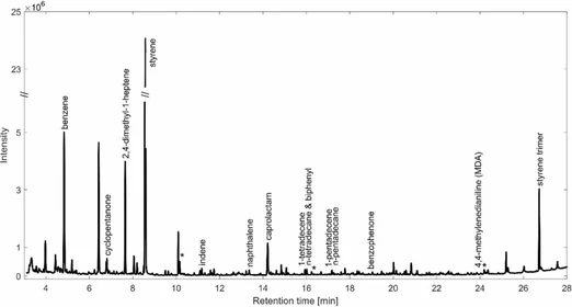
TD-GC-ICP-MS analyses were able to detect multiple organometallic species in the e-cigarette aerosol sample administered to exposed mice. Specifically, species of Al, Ni, Cu, As, Br, Sn, and Hg were detected. The chromatographic profiles demonstrated several distinct peaks corresponding to a number of metal species in aerosol samples, which were absent or negligible in air blank controls, indicating their origin from the e-cigarette device or e-liquid components (Fig. 1 and Figure S1).
 Anal Bioanal Chem 418, 3821–3835 (2026): Figure 1: Representative chromatograms of organometallic species detected in e-cigarette aerosol. A Chromatographic separation of Al, Ni, Cu, As, Br, Sn, and Hg species in aerosol samples from a Kangertech e-cigarette device (n = 3 replicates). B Corresponding air blank TD Tenex tube analysis showing the absence of these species. Analyses were performed using TD-GC-ICP-MS
As outlined above, attempts to assign molecular identities to these metal species using complementary GC-MS and GC×GC-TOF-MS were not successful. In addition, retention time comparison with available analytical standards analysed under TD-GC-ICP-MS conditions did not yield matching chromatographic features. The absence of both correspondence spectra and retention time alignment likely reflects the inherent chemical complexity, low concentrations, and thermal instability of organometallic species, as well as the limitations of current analytical approaches for their characterisation [46].
TD-GC-ICP-MS provides element-specific detection independent of molecular ionisation, enabling sensitive detection of metal-containing chromatographic entities at trace levels [46]. While the use of direct thermal desorption allows rapid analysis without derivatization or extensive sample preparation, it limits the ability to perform compound-specific speciation required for unambiguous structural or oxidation state identification. As such, detected species are reported as metal-containing chromatographic entities rather than fully characterised molecular compounds. Definitive identification would require further investigation using orthogonal high-resolution or targeted approaches (e.g. GC-Orbitrap or targeted derivatisation strategies), which were beyond the scope of this study [47]. Nevertheless, the reproducible detection of element-specific chromatographic peaks provides clear evidence for the presence of volatile or semi-volatile metal-containing species in the aerosol phase.
Elemental bioimaging of murine lung tissue following e-cigarette aerosol exposure
Lung tissue samples from control (0), 8, 16, and 32 puffs exposure groups were analysed for Cr, Fe, Ni, Cu, Zn, Sn, and Pb using LA-ICP-MS.
Vape exposure significantly reduced Fe concentrations, with both mean (p = 0.0009) and maximum (p = 0.005) values decreasing across exposure groups, although these patterns were not strictly dose-dependent. Mean Fe levels declined from 4.18 × 10³ ng g⁻1 in control lungs to 667, 597, and 1.02 × 10³ ng g⁻1 in the 8, 16, and 32 puff groups, respectively. A representative image of the Fe distribution in both the control (0 puff) and exposed group (32 puffs) is shown in Fig. 2A.
 Anal Bioanal Chem 418, 3821–3835 (2026): Fig. 2: Elemental distribution and quantification of metal accumulation in murine lungs following e-cigarette aerosol exposure. A LA-ICP-MS elemental distribution maps of targeted metals in murine lung tissue after a 4-day exposure protocol. Control lung sections (0 puffs) and aerosol-exposed sections (32 puffs) show spatial distribution and relative concentrations (ng g⁻1) of Cr, Fe, Ni, Cu, Zn, Sn, and Pb. Notable accumulation is observed in exposed tissues, indicating aerosol-associated metal deposition. B Quantitative analysis of lung tissue concentrations of Cr, Fe, Ni, Cu, Zn, Sn, and Pb across exposure groups (0, 8, 16, and 32 puffs). Fe levels significantly increased in both mean (p = 0.0009) and maximum (p = 0.005) concentrations. Ni showed a significant rise in mean concentration (p = 0.0055), while a significant effect of aerosol exposure on maximum Pb concentrations was observed (p = 0.0390). Box plots show the full data range (minimum to maximum), with individual data points overlaid. Asterisks indicate statistically significant differences from post hoc testing: *p < 0.05, **p ≤ 0.0055. C Representative elemental bioimaging of Pb accumulation in murine lungs following exposure to 8, 16, and 32 puffs administered twice daily over 4 days
Mean nickel concentrations increased significantly with exposure (p = 0.0055), rising from 77.3 ng·g⁻1 in controls to 368 ng·g⁻1 and 242 ng·g⁻1 in the 8 and 32 puff groups, respectively. Copper and lead exhibited similar trends, with maximum concentrations reaching 8.74 × 10⁴ ng·g⁻1 for Cu and 9.91 × 10⁴ ng·g⁻1 for Pb in the 32 puff group, representing 3.08-fold and 25.3-fold increases compared to control tissue samples. A significant effect of aerosol exposure on maximum Pb concentrations was observed (p = 0.0390) (Fig. 2B).
A trend was also observed where the concentrations of Sn were elevated in the 32 puff group, reaching 3.96 × 10⁵ ng·g⁻1, corresponding to a 15.4-fold increase compared to controls. Notably, Sn exhibited focal accumulation within the superior lobe, limiting statistical significance in whole-tissue comparisons. This localised deposition pattern was similarly observed for Cr, Ni, and Pb, suggesting region-specific retention of inhaled metals. In the representative images for the elements Cr, Ni, Zn, Sn, and Pb, deposits can be seen in the superior lobe (upper sections) and the pleural cavity of the lung tissue (Fig. 2B).
Representative elemental maps of lead distribution across exposure levels (8, 16, and 32 puffs) are shown in Fig. 2C, illustrating progressive dose-dependent accumulation and spatial heterogeneity.
High concentrations of Cr, Ni, Sn, and Pb were detected in the e-liquid sample, with corresponding organometallic species observed in the aerosol, and elemental bioimaging confirming their deposition in murine lung tissues. LA-ICP-MS images (Fig. 1C) showed region-specific accumulation, with Pb, Ni, and Sn concentrated in the superior lobe and pleural regions, while Zn was predominant in basilar areas. It is plausible that metals are transported to the lung via non-volatile pathways, including association with aerosol droplets, particulate matter, or nanoparticles generated during device heating. The observed spatial variation in metal deposition may likely reflect differences in particle size and physicochemical form. Aerosol deposition is governed by mechanisms such as inertial impaction, sedimentation, and diffusion, which determine regional deposition based on aerodynamic diameter. Larger particles (> 5 µm) deposit in the upper airways, whereas smaller particles (< 2 µm) reach the alveolar region, with intermediate particles depositing in the central airways [68]. In addition, these patterns may not solely reflect differences in initial deposition but also region-specific clearance processes. While particles may deposit throughout the lung, more efficient mucociliary clearance in the upper airways and slower macrophage-mediated clearance in the alveolar region could lead to differential retention and accumulation of metals across lung compartments [69]. It should also be noted that Zn is an endogenous element in lung tissue, and measured levels therefore reflect both baseline physiological concentrations and potential exposure-related contributions, limiting definitive source attribution. These heterogeneous patterns may underlie patchy lung injury and provide guidance for translational studies of vaping-related disease [70].
Conclusion
Using a multi-platform analytical approach, we identified toxic metals in e-cigarette aerosols and confirmed their focal deposition in murine lung tissue. Further, the detection of element-specific chromatographic peaks by TD-GC-ICP-MS provides clear evidence for the presence of volatile or semi-volatile metal-containing species in the aerosol phase.
The observed reduction in pulmonary iron, together with accumulation of Pb, Ni, Cu, Sn, and As, indicates disruption of metal homeostasis with potential for both local and systemic toxicity. This is the first study to apply TD-GC-ICP-MS for aerosol speciation in this context, providing insight into vaping-related risks.
Importantly, our preliminary exposure study demonstrates that even short-term vaping can result in measurable metal accumulation in lung tissue. These findings justify further investigation, including studies in human populations, to determine the broader health implications and to inform future toxicological assessments and regulatory strategies.